电极安放位置的选择是表面肌电测试的第1步,也是决定测试信号是否可靠的重要环节,因此必须高度重视。
首先,必须结合运动项目特征选取发挥主要作用的肌肉。同时,需要了解所测肌肉的主要功能和解剖学标记点(图2-4-6),这些点对电极安放很有帮助。为此,本节以European Recommendations for Surface ElectroMyo Graphy(SENIAM)为蓝本做介绍。
其次,需要了解人体浅表的主要工作肌群的名称及肌纤维的走向(决定电极粘贴方向),如图2-4-7所示(转引自The ABC of EMG[1])。

图2-4-5 采用金属丝针电极测试跑台上步行时胫骨肌的放电变化

图2-4-6 人体主要解剖学标记点

图2-4-7 浅表肌肉群的名称及电极安放位置示意图
1.上肢主要肌肉
1)肱二头肌(biceps brachii)
●组成:长头和短头(long and short head)。
●起点:长头:肩胛骨喙突点(long head:supraglenoid tubercle of scapula);短头:肩胛骨盂上结节(short head:apex of coracoid process of scapula)。
●止点:尺骨窝。
●开始姿势:受试者坐在椅子上,肘关节弯曲成直角,前臂背面水平朝下(图2-4-8)。
●电极位置:肩峰点与尺骨窝连线的远端1/3处(electrodes need to be placed on the line between the medial acromion and the fossa cubit at 1/3from the fossa cubit)。

图2-4-8 肱二头肌标志点
●电极方向:平行于肩峰点与尺骨窝连线(in the direction of the line between the acromion and the fossa cubit)。
●检测方式:检测者一手托住受试者肘关节,一手握住腕部用力向下;受试者尽可能保持肘关节90°角屈。
2)肱三头肌(triceps brachii)
●组成:长头(long head)。
●起点:肩胛骨盂下粗隆(infraglenoid tubercle of scapula)。
●止点:鹰嘴后面(posterior surface of olecranon)。

2-4-9 肱三头肌长头标志点
●开始姿势:受试者坐在椅子上,肩关节外展约90°角,肘关节弯曲成直角,前臂前面水平朝下,5指伸直向下(图2-4-9)。
●电极位置:肩峰后脊与鹰嘴连线的1/2处向内2指宽的点(electrodes need to be placed at 50%on the line between the posterior crista of the acromion and the olecranon at 2 finger widths medial to the line)。
●电极方向:平行于肩峰后脊与鹰嘴连线(in the direction of the line between the posterior crista of the acromion and the olecranon)。
●检测方式:检测者一手托住受试者肘关节,另一手握住腕部用力向内;受试者尽可能伸展肘关节,并与之对抗。
3)肱三头肌(triceps brachii)
●组成:外侧头(lateral head)。
●起点:接近于肱骨体1/2处的外侧面和后面(lateral and posterior surfaces of proximal 1/2of body of humerus and lateral intramuscular septum)。
●止点:鹰嘴后面(posterior surface of olecranon process of ulna and antebrachial fascia)。
●开始姿势:受试者坐在椅子上,肩关节外展约90°角,肘关节弯曲成直角,前臂前面水平朝下,5指伸直向下(图2-4-10)。
●电极位置:肩峰后脊与鹰嘴连线的1/2处向外2指宽的点(electrodes need to be placed at 50%on the line between the posterior crista of the acromion and the olecranon at 2 finger widths lateral to the line)。
●电极方向:平行于肩峰后脊与鹰嘴连线(in the direction of the line between the posterior crista of the acromion and the olecranon)。

图2-4-10 肱三头肌外侧头标志点
●检测方式:检测者一手托住受试者肘关节,一手握住腕部用力向内;受试者尽可能伸展肘关节,并与之对抗。
4)拇短展肌(abductor pollicis brevis)
●起点:屈肌副韧带、角骨结节、舟骨结节(flexor retinaculum,tubercle of trapezium bone and tubercle of scaphoid bone)。
●止点:拇指指骨底(base of proximal phalanx of thumb,radial side,and extensor expansion)。
●开始姿势:受试者取坐姿或仰卧,保证手背稳定放置于桌面或平台上(图2-4-11)。
●电极位置:第1掌骨近端1/4处,稍偏内(slightly medial of the distal 1/4of the 1st ossa metacarpalia)。
●电极方向:平行于第1掌骨(parallel to the 1st ossametacarpalia)。
●检测方式:检测者用中指用力下压受试者的第1指骨节;受试者尽可能伸展拇指,并与之对抗。

图2-4-11 拇短展肌标志点
2.下肢主要肌肉
1)股四头肌(quadriceps femoris)
●组成:股直肌(rectus femoris)。
●起点:髂前下棘(straight head from anterior inferior iliac spine;reflected head from groove above rim of acetabulum)。
●止点:通过髌韧带,止于胫骨粗隆(proximal border of the patella and through patellar ligament)。

2-4-12 股直肌标志点
●开始姿势:受试者坐在台子上,膝关节微屈,上体后仰,小腿稍抬起(图2-4-12)。
●电极位置:髂前上棘和髌骨上部的连线50%处(the electrodes need to be placed at 50%on the line from the anterior spina iliaca superior to the superior part of the patella)。
●电极方向:与髂前上棘和髌骨上部的连线平行(in the direction of the line from the anterior spina iliaca superior to the superior part of the patella)。
●检测方式:检测者一手托住受试者膝关节,另一手握住踝部用力向下;受试者尽可能伸展膝关节,并与之对抗。
2)股四头肌(quadriceps femoris)
●组成:股内侧肌(vastus medialis)。

图2-4-13 股内侧肌标志点
●起点:股骨粗线内侧唇(distal half of the intertrochanteric line,medial lip of line aspera,proximal part of medial supracondylar line,tendons of adductor longus and adductor magnus and medial intermuscular septum)。
●止点:通过髌韧带,止于胫骨粗隆(proximal border of the patella and through patellar ligament)。
●开始姿势:受试者坐在台子上,上体后仰,膝关节微屈,小腿稍抬起(图2-4-13)。
●电极位置:髂骨前棘和髌内侧韧带关节缝连线的远端80%处(electrodes need to be placed at 80%on the line between the anterior spina iliaca superior and the joint space in front of the anterior border of the medial ligament)。
●电极方向:几乎垂直于髂骨前棘和髌内侧韧带关节缝的连线(almost perpendicular to the line between the anterior spina iliaca superior and the joint space in front of the anterior border of the medial ligament)。
●检测方式:检测者一手托住受试者膝关节,另一手握住踝部用力向下;受试者尽可能伸展膝关节,并与之对抗。
3)股四头肌(quadriceps femoris)
●组成:股外侧肌(vastus lateralis)。
●起点:股骨粗线外侧唇,接近大转子线(proximal parts of intertrochanteric line,anterior and inferior borders of greater trochanter,lateral lip of gluteal tuberosity,proximal half of lateral lip of linea aspera,and lateral intermuscular septum)。
●止点:通过髌韧带,止于胫骨粗隆(proximal border of the patella and through patellar ligament)。

图2-4-14 股外侧肌标志点
●开始姿势:受试者坐在台子上,上体后仰,小腿稍抬起,膝关节微屈(图2-4-14)。
●电极位置:髂前上棘和髌骨外侧连线的远端2/3处(electrodes need to be placed at 2/3on the line from the anterior spina iliaca superior to the lateral side of the patella)。
●电极方向:与髂前上棘和髌骨外侧的连线平行(in the direction of the muscle fibres)。
●检测方式:检测者一手托住受试者膝关节,另一手握住踝部用力向下;受试者尽可能伸展膝关节,与之对抗。
4)半腱肌(semitendinosus)
●起点:坐骨结节(tuberosity of ischium by tendon common with long head of biceps femoris)。
●止点:胫骨粗隆内侧面(proximal part of medial surface of body of tibia and deep fascia of leg)。

图2-4-15 半腱肌标志点
●开始姿势:受试者俯卧在台子上,膝关节微屈,小腿稍抬起(图2-4-15)。
●电极位置:坐骨结节和胫骨中间骨节连线的50%处(electrodes need to be placed at 50%on the line between the ischial tuberosity and the medial epycondyle of the tibia)。
●电极方向:与坐骨结节和胫骨中间骨节的连线平行(in the direction of the line between the ischial tuberosity and the medial epycondyle of the tibia)。
●检测方式:检测者一手托住受试者踝关节,另一手按住臀部用力向外;受试者尽可能屈膝关节,并与之对抗。
5)股二头肌(biceps femoris)
●组成:长头。
●起点:长头起自坐骨结节,短头起自股骨粗线外侧唇上半部(long head:distal part of sacrotuberous ligament and posterior part of tuberosity)。
●止点:腓骨头(lateral side of head of fibula,lateral condyle of tibia,deep fascial on lateral side of leg)。
●开始姿势:受试者取俯卧位,大腿与台子贴近,小腿稍抬起,膝关节弯曲约90°角(图2-4-16)。
●电极位置:坐骨结节与胫骨外侧髁连线的50%处(the electrodes need to be placed at 50%on the line between the ischial tuberosity and the lateral epicondyle of the tibia)。
●电极方向:与坐骨结节和胫骨外侧髁的连线平行(in the direction of the line between the ischial tuberosity and the lateral epicondyle of the tibia)。

图2-4-16 肌二头肌长头标志点
●检测方式:检测者一手托住受试者踝关节,另一手按住臀部用力向外;受试者尽可能屈膝关节,并与之对抗。
6)胫骨前肌(tibialis anterior)
●起点:胫骨体外侧的上2/3(lateral condyle and proximal 1/2of lateral surface of tibia,interosseus membrane,deep fascia and lateral intermuscular septum)。
●止点:肌腱从内踝前方通过,止于内侧(第1)楔骨和第1跖骨底(medial and plantar surface of medial cuneiform bone,base of first metatarsal bone)。

2-4-17 胫骨前肌标志点
●开始姿势:受试者取坐姿,上体略后仰,膝关节微弯曲,踝关节可以背屈(图2-4-17)。
●电极位置:腓骨头顶端和踝关节内侧髁连线的近端1/3处(the electrodes need to be placed at 1/3on the line between the tip of the fibula and the tip of the medial malleolus)。
●电极方向:与腓骨头顶端和踝关节内侧髁的连线平行(in the direction of the line between the tip of the fibula and the tip of the medial malleolus)。
●检测方式:检测者一手扶住受试者膝关节,另一手用力向下按压脚面;受试者尽可能背屈踝关节(勾脚尖),并与之对抗。

图2-4-18 腓肠肌外侧头标志点
7)腓肠肌(gastrocnemius)
●组成:外侧头(lateralis)。
●起点:股骨外上髁和股骨后面(lateral condyle and posterior surface of femur,capsule of knee joint)。
●止点:跟骨结节(middle part of posterior surface of calcaneus)。
●开始姿势:受试者取俯卧位,大腿贴于台面,小腿略抬起(图2-4-18)。
●电极位置:跟骨与腓骨头连线近端的1/3处(electrodes need to be placed at 1/3of the line between the head of the fibula and the heel)。
●电极方向:与跟骨及腓骨头的连线平行(in the direction of the line between the head of the fibula and the heel)。
●检测方式:受试者尽可能伸展踝关节,并与检测者对抗。
8)腓肠肌(gastrocnemius)
●组成:内侧头(medialis)。
●起点:股骨内上髁(proximal and posterior part of medial condyle and adjacent part of the femur,capsule of the knee joint)。
●止点:跟骨结节(middle part of posterior surface of calcaneus)。

2-4-19 腓肠肌内侧头标志点
●开始姿势:受试者取俯卧位,大腿贴于台面,小腿略抬起(图2-4-19)。
●电极位置:放置在最突出鼓起的肌腹处(electrodes need to be placed on the most prominent bulge of the muscle)。
●电极方向:与胫骨平行(in the direction of the leg)。
●检测方式:受试者尽可能伸展踝关节,并与检测者对抗。
9)腓骨短肌(peroneus brevis)
●起点:腓骨的外侧面(distal 2/3of lateral surface of fibula and adjacent intermuscular septa)。
●止点:腱经外踝的后面转向前,止于第5跖骨粗隆(tuberosity at base of fifth metatarsal bone,lateral side)。
●开始姿势:坐姿,上体略后仰,膝关节微弯曲,踝关节外翻(图2-4-20)。
●电极位置:腓骨头和腓骨外侧髁连线,远端1/4处(electrodes need to be placed anterior to the tendon of the m.peroneus longus at 25%of the line from the tip of the lateral malleolus to the fibula-head)。
●电极方向:平行于腓骨头和腓骨外侧髁连线(in the direction of the line from the tip of the lateral malleolus to the fibula-head)。

图2-4-20 胫骨短肌标志点
●检测方式:受试者踝关节外翻,检测者用手与之对抗。
10)腓骨长肌(peroneus longus)
●起点:腓骨体的外侧面,肌腱经外踝后面转至足底(lateral condyle of tibia,head and proximal 2/3of lateral surface of fibula,intermuscular septa,and adjacent deep fascia)。
●止点:内侧楔骨及第1跖骨底和第5跖骨粗隆(lateral side of base of first metatarsal and of medial cuneiform bone)。
●开始姿势:受试者取坐姿,上体略后仰,膝关节微弯曲,踝关节外翻(图2-4-21)。
●电极位置:腓骨头和外侧髁连线近端1/4处(electrodes need to be placed at 25%on the line between the tip of the head of the fibula to the tip of the lateral malleolus)。
●电极方向:与腓骨头和外侧髁连线平行(in the direction of the line between the tip of the head of the fibula to the tip of the lateral malleolus)。
●检测方式:受试踝关节外翻,检测者用手与之对抗。
11)比目鱼肌(soleus)
●起点:胫骨和腓骨后上部的比目鱼肌线(posterior surfaces of the head of the fibula and proximal 1/3of its body,soleal line and middle 1/3 of medial border of tibia and tendinous arch between tibia and fibula)。
●止点:跟骨结节(with tendon of gastrocnemius into posterior surface of calcaneus)。

图2-4-21 腓骨长肌标志点
●开始姿势:受试者取坐姿,屈膝将脚平放在台面上(图2-4-22)。
●电极位置:胫骨头内侧面中心点到踝内侧髁连线的远端2/3处(the electrodes need to be placed at 2/3of the line between the medial condylis of the femur to the medial malleolus)。
●电极方向:与胫骨头内侧面中心点到踝内侧髁的连线平行(in the direction of the line between the medial condylis to the medial malleolus)。

2-4-22 比目鱼肌标志点
●检测方式:检测者双手按住受试者膝关节;受试者尽可能伸展踝关节,并与之对抗。
3.髋部主要肌肉群
1)臀肌(gluteus)
●组成:臀大肌(maximus)。
●起点:髂骨翼外面及骶、尾骨背面(posterior gluteal line of ilium ad portion of bone superior and posterior to the posterior surface of lower part of sacrum,side of coccyx,aponeurosis of erector spinea,sacrotuberous ligament and gluteal aponeurosis)。
●止点:股骨臀肌粗隆和髂胫束(larger proximal portion and superficial fibres of distal portion of muscle into iliotibial tract of fascia lata;deeper fibres of distal portion into gluteal tuberosity of femur)。

图2-4-23 臀大肌标志点
●开始姿势:受试者取俯卧在测试台上(图2-4-23)。
●电极位置:骶椎和大转子连线的50%处,正好处于大转子上方臀中部最凸出的肌肉上(the electrodes need to be placed at 50%on the line between the sacral vertebrae and the greater trochanter.This position corresponds with the greatest prominence of the middle of the buttocks well above the visible bulge of the greater trochanter)。
●电极方向:从髂后上棘到大腿后面的方向(in the direction of the line from the posterior superior iliac spine to the middle of the posterior aspect of the thigh)。
●检测方式:检测者用力按住踝关节向下用力;受试者尽可能伸髋关节,并与之对抗。
2)臀肌(gluteus)
●组成:臀中肌(medius)。
●起点:髂骨翼外面(external surface of ilium between iliac crest and posterior gluteal line dorsally,and anterior gluteal line ventrally,gluteal aponeurosis)。
●止点:股骨大转子外侧面斜嵴(oblique ridge on lateral surface of greater trochanter of femur)。

图2-4-24 臀中肌标志点
●开始姿势:受试者侧卧(图2-4-24)。
●电极位置:髂嵴到大转子的1/2处(electrodes need to be placed at 50%on the line from the crista iliaca to the trochanter)。
●电极方向:髂嵴到大转子连线平行(in the direction of the line from the crista iliaca to the trochanter)。
●检测方式:令受试者的大腿做屈伸、旋内、旋外。
3)阔筋膜张肌(tensor fasciae latae)
●起点:髂前上棘(anterior part of external lip of iliac crest,outer surface of anterior superior iliac spine and deep surface of fascia lata)。
●止点:该肌在大腿外侧移行于髂胫束,止于胫骨外侧髁(into iliotibial tract of fascia lata at junction of proximal and middle thirds of the thigh)。

图2-4-25 阔筋膜张肌标志点
●开始姿势:受试者侧卧于实验台上,膝关节微屈,大腿外展(图2-4-25)。
●电极位置:髂前上棘与胫骨外侧髁连线近端1/6处(on the line from the anterior spina iliaca superior to the lateral femoral condyle in the proximal 1/6)。
●电极方向:与髂前上棘与胫骨外侧髁的连线平行(in the direction of the line from the anterior spina iliaca superior to the lateral femoral condyle)。
●检测方式:检测者一手抵住受试者膝关节,另一手扶住髋部;受试者尽可能屈髋关节外展,并与之对抗。

2-4-26 三角肌前部标志点
4.肩背部主要肌群
1)三角肌(deltoideus)
●组成:前部(anterior)。
●起点:锁骨外侧1/3、肩峰和肩胛冈(anterior border,superior surface,lateral 1/3of clavicle)。
●止点:肱骨体三角肌粗隆(deltoid tuberosity of humerus)。
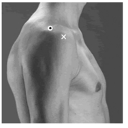
●开始姿势:受试者直立(图2-4-26)。
●电极位置:肩峰前部1个手指宽的位置上(the electrodes need to be placed at one finger width distal and anterior to the acromion)。
●电极方向:肩峰到拇指的方向(in the direction of the line between the acromion and the thumb)。
●检测方式:检测者一手托住受试者肩关节后部,另一手握住腕部用力向后;受试者尽可能直臂前摆,并与之对抗。
2)三角肌(deltoideus)
●组成:后部(posterior)。
●起点:肩胛冈的后缘(inferior lip of posterior border of spine of scapula)。
●止点:肱骨体三角肌粗隆(deltoid tuberosity of humerus)。
●开始姿势:受试者直立(图2-4-27)。
●电极位置:肩峰角后两个手指宽的位置(center the electrodes in the area about two finger breaths behind the angle of the acromion)。
●电极方向:从肩峰到小拇指方向(in the direction of the line between the acromion and the little finger)。
●检测方式:检测者一手托住受试者肩关节前部,另一手握住腕部用力向前;受试者尽可能直臂后摆,并与之对抗。
3)三角肌(deltoideus)
●组成:中部(medius)。
●起点:肩峰上缘和侧缘(lateral margin and superior surface of the acromion)。

图2-4-27 三角肌后部标志点
●止点:肱骨体三角肌粗隆(deltoid tuberosity of humerus)。
●开始姿势:受试者直立(图2-4-28)。
●电极位置:肩峰到肘关节外侧髁连线,肌肉最鼓起的部分(electrodes need to be placed from the acromion to the lateral epicondyle of the elbow;this should correspond to the greatest bulge of the muscle)。
●电极方向:从肩峰到手的方向(in the direction of the line between the acromion and the hand)。
●检测方式:检测者一手扶住受试者肘关节,另一手握住腕部用力向内;受试者尽可能外展肩关节,并与之对抗。

2-4-28 三角肌中部标志点
4)斜方肌(trapezius)
●组成:上部[trapezius descendens(upper)]
●起点:枕外隆凸、项韧带及颈椎骨第7棘突(external occipital protuberance,medial 1/3of superior nuchal line,ligamentum nuchae,and spinous process of 7th cervical vertebra)。
●止点:锁骨外侧1/3、肩峰(lateral 1/3of clavicle and acromion process of scapula)。
●开始姿势:受试者直立(图2-4-29)。

图2-4-29 斜方肌上部标志点
●电极位置:肩峰到第7颈椎连线的50%处(the electrodes need to be placed at 50%on the line from the acromion to the spine on vertebra C7)。
●电极方向:与肩峰和第7颈椎的连线平行(in the direction of the line between the acromion and the spine on vertebra C7)。
●检测方式:受试者尽可能提肩。
5)斜方肌(trapezius)
●组成:中部[transversalis(middle)]。
●起点:第1~5胸椎棘突(spinous processes of first through fifth thoracic vertebrae)。
●止点:肩胛冈边缘(superior lip of spine of scapula)。
●开始姿势:受试者直立(图2-4-30)。

2-4-30 斜方肌中部标志点
●电极位置:肩胛骨中部边缘线和第3胸椎棘突连线50%处(the electrodes need to be placed at 50%between the medial border of the scapula and the spine,at the level of T3)。
●电极方向:肩峰到第5胸椎(in the direction of the line between T5and the acromion)。
●检测方式:受试者做扩胸飞鸟动作。
6)斜方肌(trapezius)
●组成:下部[trapezius ascendens(lower)]。
●起点:第6到第12胸椎棘突(spinous processes of sixth to twelfth thoracic vertebrae)。
●止点:肩胛冈顶点(apex of spine of scapula)。
●开始姿势:受试者直立(图2-4-31)。
●电极位置:从肩胛骨三角区域到第8胸椎连线的远端2/3处(the electrodes need to be placed at 2/3on the line from the trigonum spinea to the 8th thoracic vertebra)。
●电极方向:从肩峰到第8胸椎(in the direction of the line between T8and the acromion)。

图2-4-31 斜方肌下部标志点
●检测方式:受试者做扩胸飞鸟动作。
5.腰部主要肌群(reference electrode:on the proc spin of C7)
1)竖脊肌(erector spinae)
●组成:最长肌(longissimus)。
●起点:骶骨背面和髂嵴后部(in lumbar region is blended with iliocostalis lumborum,posterior surfaces of transverse and accessory processes of lumbar vertebrae,and anterior layer of thoracolumbar fascia)。
●止点:椎骨横突,上到达颞骨乳突(by tendons into tips of transverse process of all thoracic vertebrae,and by fleshy digitations anto lower 9or 10ribs between tubercles and angles)。
●开始姿势:受试者直立(图2-4-32)。
●电极位置:第1腰椎外侧2手指宽处(the electrodes need to be placed at 2finger width lateral from the proc.spin.of L1)。
●电极方向:参考解剖标志点图,电极上下垂直(vertical)。
●检测方式:受试者俯卧抬上体背伸对抗。
2)棘肌(multifidus)
●起点:椎骨棘突(spinous processes L1~L5)。
●止点:骶骨背面和髂嵴后部(massillary processes L4~S1 and iliaccresr,dorsal surface of sacrum)。
●开始姿势:受试者直立(图2-4-33)。

图2-4-32 竖脊肌标志点
●电极位置:在第1和第2腰椎连接缝与后髂棘突连线中心下2~3cm处,位于第5腰椎水平(electrodes need to be placed on and aligned with a line from caudal tip posterior spina iliaca superior to the interspace between L1and L2interspace at the level of L5spinous process(i.e.about 2-3cm from the midline)。
●电极方向:与第1和第2腰椎连接缝与后髂棘突连线平行(in the direction of the line described above)。
●检测方式:受试者俯卧,抬上体,背伸对抗。

图2-4-33 棘肌标志点
3)髂肋肌(iliocostalis)
●起点:骶骨背面和髂嵴后部(anterior surface of broad tendon attached to medial crest of sacrum,spinous processes of lumbar and 11th and 12th thoracic vertebrae,posterior part of medial lip of iliac crest,supraspinous ligament and lateral crest of sacrum)。
●止点:第6或第7肋骨下缘(by tendons into inferior border of angles of lower 6or 7 ribs)。
●开始姿势:受试者直立(图2-4-34)。
●电极位置:在后髂骨棘突末端和肋骨下缘连线外侧1指宽,与第2腰椎平齐(the electrodes need to be placed 1finger width medial from the line from the posterior spina iliaca superior to the lowest point of the lower rib,at the level of L2)。
●电极方向:后髂骨棘突末端和肋骨下缘连线平行(in the direction of the line between the posterior spina iliaca superior and lowest point of the lower rib)。
●检测方式:受试者俯卧,抬上体,背伸对抗。
《康复医学肌电使用指导手册》(Surface emg made easy:a beginner's guide for rehabilitation clinicians[2])中显示了建议的电极和参考电极的位置。电极粘贴组图如(图2-4-35)。

图2-4-34 髂肋肌标志点


图2-4-35 《康复医学肌电使用指导手册》中建议的各块肌肉的电极和参考电极的安放位置
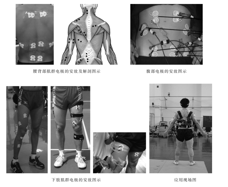
以上组图是文献中提供的,供读者在肌肉功能障碍诊断时参考,特别是一些不常用的肌肉群以及股四头肌、腘绳肌等肌群组的测试方式值得借鉴。图中独立电极(黑色)为参考电极(地线),沿肌肉方向放置的配对电极为测试电极。图2-4-36是腰背部及下肢等肌群电极的安放图示。图2-4-37是Mega6000表面肌电仪自带软件Megawin中的肌肉图谱标示。

图2-4-36 各肌群电极安放图示


图2-4-37 Mega6000表面肌电仪自带软件Megawin中的肌肉图谱标示
免责声明:以上内容源自网络,版权归原作者所有,如有侵犯您的原创版权请告知,我们将尽快删除相关内容。


