第八节 共聚焦显微镜在其他角膜病中的应用
一、干眼症
干眼症亦称角结膜干燥症,是极为常见的眼表疾病,是指任何原因引起的泪液质和量或动力学异常导致的泪膜不稳定,并伴有眼部不适症状,眼表组织病变为特征的一大类疾病的总称(图4-75)。如仅有眼部不适症状而无眼表损伤的体征,称为干眼症。如同时伴有眼表损伤,称为干眼病。

图4-75 干眼症患者的裂隙灯像
注:角膜上皮点状染色,泪河变浅
在共聚焦显微镜下,干眼症患者一般表现为眼表各层上皮细胞特别是鳞状上皮细胞增生,上皮细胞呈应激状态,细胞核突出;部分患者可伴有上皮下神经改变,如神经增粗、分支增多、走行不规则等。这些均为干眼症在共聚焦显微镜下的常见表现(图4-76)。

A.角膜浅表鳞状上皮细胞增生,排列密集,细胞核明显突出,细胞呈激活状态,并可见胞体高亮的脱落细胞(箭头)

B.角膜基底层上皮细胞胞体增大,细胞核突出(箭头),细胞呈激活状态

C.角膜基底层上皮细胞间隙增大,细胞核突出(箭头),细胞呈激活状态

D.角膜上皮(下方)-基质(上方)交界处可见上皮下神经明显增粗,走行尚规则

E.角膜上皮下神经明显增粗,排列密集,伴少量朗格汉斯细胞浸润(箭头)

F.角膜上皮下神经明显增粗,伴少量朗格汉斯细胞浸润(箭头)

G.角膜上皮下神经明显增粗,伴少量朗格汉斯细胞浸润(箭头)
图4-76 共聚焦显微镜观察干眼症患者角膜(A~D为X 1 000, NIDEK CS系列;E~F为X 800, HRT系列)
二、眼类天疱疮
眼类天疱疮可导致黏蛋白缺乏性干眼病。本病常隐匿发病,有眼红、异物感、流泪、畏光等症状。双侧发病,病程反复。典型体征为下方睑球粘连、下穹窿缩窄(图4-77)。

A.角膜广泛新生血管,中央角膜基质部分溶解

B.可见广泛结膜瘢痕
图4-77 眼类天疱疮患者的裂隙灯像
在共聚焦显微镜下,患者角膜除存在角膜各层上皮细胞增生、激活的干眼病共同表现外,还可见角膜、结膜炎症细胞浸润,并常累及基质。基质细胞呈激活状态,有时可见基质瘢痕形成。由于角膜基质层透光性下降,内皮细胞形态大多较模糊(图4-78)。

A.结膜内见大量炎症细胞浸润

B.角膜上皮纵切面,可见眼表各层上皮细胞特别是表层鳞状上皮细胞增生,各层上皮细胞呈激活状态,细胞核突出

C.角膜翼状细胞胞体增大,细胞核突出(箭头),细胞呈激活状态

D.角膜基底细胞增生,细胞密集,细胞核突出(黑色箭头),细胞呈激活状态,并可见少量圆形高反光炎症细胞浸润(白色箭头)

E.基质细胞呈明显激活状态,胞体相互交织呈致密网状
图4-78 共聚焦显微镜观察眼类天疱疮患者的角膜(X 800,HRT系列)
三、Sjögren综合征
Sjögren综合征是一组由自身免疫性疾病引起的综合征。除了干眼症的眼部表现外,还包括角结膜干燥,口腔、鼻腔及生殖道黏膜干燥和结缔组织病。
Sjögren综合征多发生于绝经期妇女,眼部主要表现为一系列干眼症状,但病情严重程度重于普通的干眼症患者(图4-79)。 Sjögren综合征的病理检查表现以泪腺中大量淋巴细胞浸润、泪液分泌功能破坏为特征,可致泪液生成不足性干眼病。病变晚期,腺泡组织被大量的纤维结缔组织所取代。

图4-79 Sjögren患者的裂隙灯像注:角膜荧光染色后可见角膜上皮广泛糜烂
在共聚焦显微镜下,Sjögren综合征患者除存在角膜各层上皮细胞增生、激活等的干眼病共同表现外,还可见角膜、结膜炎症细胞浸润,并常累及基质。基质细胞呈激活状态,有时可见前弹力层和基质沉积物(图4-80)。

A.角膜浅表上皮细胞增生,细胞核突出(黑色箭头),细胞呈激活状态;并可见少量圆形高反光炎症细胞浸润(白色箭头)

B.角膜基底层上皮细胞增生,细胞核突出(黑色箭头),细胞呈激活状态;并可见少量圆形高反光炎症细胞浸润(白色箭头)

C.角膜基底层上皮细胞排列紊乱,细胞核突出,细胞呈激活状态。基底膜反光增强,局部皱褶(箭头)

D.角膜上皮基底膜可见不规则形态高反光沉积物(白色箭头),上皮下神经纤细,走行紊乱,可见断端(黑色箭头)

E.角膜基质细胞呈明显激活状态,胞体相互交织呈致密网状

F.角膜基质混浊,背景反光较强,基质细胞间可见细针状高反光沉积物(箭头)
图4-80 共聚焦显微镜观察Sjögren患者的角膜(X 800,HRT系列)
四、类风湿关节炎伴发干眼病
类风湿关节炎是常见的全身性结缔组织病,常伴发干眼病。共聚焦显微镜下,患者除存在角膜各层上皮细胞增生、激活等的干眼病共同表现外,还常伴有上皮下神经数量增多、减少、分支增多、走行紊乱等,有时可见前弹力层和浅基质层沉积物、瘢痕(图4-81)。

A.角膜基底细胞增生,细胞密集,细胞核明显突出,细胞呈激活状态

B.上皮下神经明显增粗,数量增多,分支增多,相互交联。不伴明显炎症细胞浸润

C.上皮下神经分支明显增多,相互交联呈网状
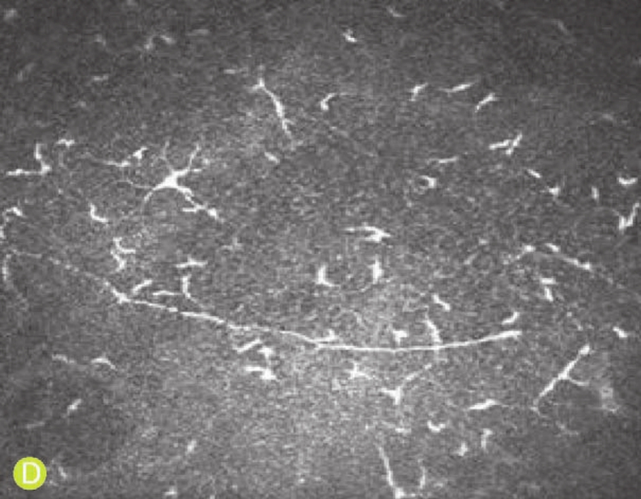
D.上皮下神经明显减少,未见分支,伴中量朗格汉斯细胞浸润

E.前弹力层界面高反光瘢痕组织(箭头),瘢痕处神经走行中止

F.患者对侧眼前弹力层界面高反光瘢痕组织(箭头),瘢痕处神经走行中止

G.患者对侧眼浅基质层,可见高反光瘢痕组织(箭头)
图4-81 共聚焦显微镜观察类风湿关节炎伴干眼病患者的角膜
(X 800,HRT系列)
五、丝状角膜病变
干眼病可引起丝状角膜病变。患者局部角膜上皮细胞卷成丝状物,一端附在角膜表面,另一端呈游离状态。自觉症状有异物感及眼痛。裂隙灯下可见角膜表面挂有细丝状物,以上方角膜较多见(图4-82)。共聚焦显微镜下可见多枚脱落的角膜上皮细胞卷成一高反光条索,顶端悬浮,末端连于角膜上皮层内(图4-83)。

A.角膜上皮呈丝状剥脱,附着于角膜表面

B.钴蓝光下卷丝清晰可见(箭头处)
图4-82 丝状角膜病变患者的裂隙灯像

图4-83 共聚焦显微镜观察丝状角膜病变患者的角膜(X 800,HRT系列)
注:多枚脱落的角膜上皮细胞卷成一高反光条索,顶端悬浮,末端连于角膜上皮层内(箭头)
(乐琦骅 徐建江 洪佳旭 郑天玉 朱文卿)
参考文献
1.王琪,陈家祺,肖迎,等.共聚焦显微镜在角膜营养不良诊断中的应用。眼科, 2005, 14(3):172~175
2.史伟云,谢立信,李绍伟,等.圆锥角膜患者表面角膜镜片术后镜片细胞和神经重建的活体动态观察.中华眼科杂志, 2002, 38(5):295~297
3.史伟云,华晓光,王富华,等.真菌性角膜炎药物治疗后转归的共聚焦显微镜观察.中华眼科杂志, 2005, 41(7):614~619
4.孙旭光,庞国祥,王智群,等.共聚焦显微镜诊断棘阿米巴性角膜炎2例.中华眼科杂志, 1999, 35(5):400~401
5.乐琦骅,徐建江.深板层角膜移植术后全层角膜组织的共聚焦显微镜观察.中华眼科杂志, 2007, 43(10):936~939
6.刘广峰,洪晶.深板层与穿透性角膜移植术后角膜内皮细胞密度的比较研究.中国实用眼科杂志, 2005, 23(9):921~924
7.李绍伟, Gebhardt BM,史伟云,等.共聚焦显微镜鉴别诊断真菌性角膜炎的实验研究.眼科研究, 2001, 19(5):389~392
8.李航,王立,邹留河,等.共聚焦显微镜在棘阿米巴性角膜炎临床诊断中的应用.眼科, 2003, 12(6):336~338
9.李绍伟, Beue RW.利用共聚焦显微镜对穿透角膜移植术后散光的研究.眼科研究, 2000, 18(5):436~438
10.李莉,何世坤.板层角膜移植术.眼科研究, 2002, 20(4):370~372
11.张琛,孙旭光.共聚焦显微镜诊断双眼角膜塑型镜相关性阿米巴角膜炎1例.眼视光学杂志, 2007, 3:182~187
12.张军,王丽娅,孙声桃,等.镰孢菌属真菌性角膜炎的临床及共聚焦显微镜特征分析.眼科研究, 2008, 26(3):219~221
13.肖启国,刘祖国,罗丽辉,等.颗粒状角膜营养不良活体共聚焦显微镜形态学研究.中国实用眼科杂志, 2006, 24(4):395~398
14.肖启国,罗丽辉,陈媛,等.早期圆锥角膜共聚焦显微镜形态学研究.南华大学学报(医学版), 2006, 34(2):168~171
15.罗丽辉,刘祖国,陈龙山,等.角膜移植术后角膜在共聚焦显微镜下的形态学改变.眼科学报, 2003, 19:201~205
16.罗丽辉,刘祖国,张梅,等.圆锥角膜患者活体共聚焦显微镜的影像改变.中华眼科杂志, 2005, 41(7):656~658
17.林跃生,孙明霞,陈家祺,等.角膜移植排斥反应的共聚焦显微镜研究.中国实用眼科杂志, 2001, 19(8):592~595
18.郑彩慧,朱学军.共聚焦显微镜在角膜营养不良中的应用.医学综述, 2008, 14(2):30~303
19.郭宁,周跃华,瞿佳,等.准分子激光原位角膜磨镶术后弥漫性板层角膜炎的共聚焦显微镜观察.中华眼科杂志, 2006, 42(4):330~333
20.洪晶,刘广峰,夏宁,等.小切口下角膜后弹力层剥除联合深板层内皮移植术的实验研究.中华眼科杂志, 2008, 44(2):122~127
21.姚勇,刘祖国,陈龙山,等.圆锥角膜对侧无明显改变眼的共聚焦显微镜所见.中国实用眼科杂志, 2005, 23(3):293~296
22.荣蓓,晏晓明.激光共聚焦显微镜观察长期佩戴软性角膜接触镜者的角膜缘改变.中华眼科杂志, 2007, 43(6):514~518
23.徐建江,洪佳旭,王艳.角膜后弹力层剥除联合内皮移植术治疗大泡性角膜病变.中华眼科杂志, 2007, 43(7):662~663
24.倪逴.眼的病理解剖与临床.上海:上海科学普及出版社, 2002. 101
25.徐丽,邹留河,李航,等.圆锥角膜各期的共聚焦显微镜表现.眼科研究, 2007, 25(4):295~298
26.黄挺,陈家祺,王玉娟,等.小切口无缝线深板层角膜内皮移植术的疗效观察.中华眼科杂志, 2007, 34(2):118~123
27.谢立信,李绍伟,史伟云,等.共聚焦显微镜在真菌性角膜炎临床诊断中的应用.中华眼科杂志, 1999, 35(1):7~9
28.韩玉萍,李冰,赵炬伟,等.激光扫描共聚焦显微镜观察穿透角膜移植术后角膜的形态学特征.中国实用眼科杂志, 2007, 25(3):306~308
29.董微丽,邹留河,潘志强,等.应用共聚焦显微镜观察Fuch角膜内皮营养不良患者的病变形态学特征.中华眼科杂志, 2004, 40(7):465~470
30.黎明,林跃生,姚晓明,等.板层角膜移植术后全层角膜组织改变的共聚焦显微镜观察.中国实用眼科杂志, 2002, 23(4):352~355
31.潘飞,张蓓,姚玉峰,等.共聚焦显微镜在临床诊断真菌性角膜炎中的应用.中国实用眼科杂志, 2004, 22(1):23~25
32. Alsuhaibani AH, Al-Rajhi AA, Al-Motowa SM, et al. Corneal endothelial cell density and morphology after acute hydrops in keratoconus. Cornea, 2008, 27(5):535~538
33. Al-Torbak AA, Al-Motowa S, Al-Assiri A, et al. Deep anterior lamellar keratoplasty for keratoconus. Cornea, 2006, 25:408~412
34. Awwad ST, Petroll WM, McCulley JP, et al. Updates in Acanthamoeba keratitis. Eye Contact Lens, 2007, 33(1):1~8
35. Babu K, Murthy KR. Combined fungal and Acanthamoeba keratitis: diagnosis by in vivo confocal microscopy. Eye, 2007, 21(2):271~272
36. Babu K, Murthy KR. In vivo confocal microscopy in different types of posterior polymorphous dystrophy. Indian J Ophthalmol, 2007, 55(5): 376~378
37. Balestrazzi A, Martone G, Traversi C, et al. Keratoconus associated with corneal macular dystrophy: in vivo confocal microscopic evaluation. Eur J Ophthalmol, 2006, 16(5):745~750
38. Barabino S, Rolando M. In vivo confocal microscopy of ocular cicatricial pemphigoid. Ophthalmic Surg Las Imag, 2006, 37(2):175~176
39. Baudouin C, Bourcier T, Dupas B, et al. Contribution of corneal in vivo confocal microscopy to the exploration of the ocular surface. Paris: Quinze-Vingts, 2006.78~84
40. Benítez del Castillo JM, Wasfy MA, Fernandez C,et al. An in vivo confocal masked study on corneal epithelium and subbasal nerves in patients with dry eye. Invest Ophthalmol Vis Sci, 2004, 45(9):3030~3035
41. Bourne WM. Cellular changes in transplanted human corneas. Cornea, 2001, 20(6):560~569
42. Brasnu E, Bourcier T, Dupas B, et al.In vivo confocal microscopy in fungal keratitis. Br J Ophthalmol, 2007, 91(5):588~591
43. Braunstein RE,Jain S, McCally RL,et al. Objective measurement of corneal light scattering after excimer laser keratectomy. Ophthalmology, 1996, 103(3):439~443
44. Bühren J, Baumeister M, Cichocki M, er al. Confocal microscopic characteristics of stage 1 to 4 diffuse lamellar keratitis after laser in situ keratomileusis. J Cataract Refract Surg, 2002, 28(8):1390~1399
45. Bühren J, Baumeister M, Kohnen T. Diffuse lamellar keratitis after laser in situ keratomileusis imaged by confocal microscopy. Ophthalmology, 2001, 108(6):1075~1081
46. Bühren J, Cichocki M, Baumeister M, et al. Diffuse lamellar keratitis after laser in situ keratomileusis. Clinical and confocal microscopy findings. Ophthalmology, 2002, 99(3):176~180
47. Cavanagh HD, Jester JV, Essepian J, et al. Confocal microscopy of the living eye. CLAO J, 1990, 16(1):65~73
48. Cavanagh HD, McCulley JP.In vivo confocal microscopy and Acanthamoeba keratitis. Am J Ophthalmol, 1996, 121(2):207~208
49. Cavanagh HD, Petroll WM, Alizadeh H, et al. Clinical and diagnostic use of in vivo confocal microscopy in patients with corneal disease. Ophthalmology, 1993, 100(10):1444~1454
50. Chen WL, Chang HW, Hu FR. In vivo confocal microscopic evaluation of corneal wound healing after epi-LASIK. Invest Ophthalmol Vis Sci, 2008, 49(6):2416~2423
51. Cheng LL, Young AL, Wong AK, et al. Confocal microscopy of posterior polymorphous endothelial dystrophy. Cornea, 2005, 24(5):599~602
52. Chew SJ, Beuerman RW, Assouline M, et al. Early diagnosis of infectious keratitis with in vivo real time confocal microscopy. CLAO J, 1992, 18(3): 197~201
53. Cho BJ, Holland EJ. In vivo tandem scanning confocal microscopy in Acanthamoeba keratitis. Korean J Ophthalmol, 1998, 12(2):112~117
54. Darwish T, Brahma A, O'Donnell C, et al. Subbasal nerve fiber regeneration after LASIK and LASEK assessed by noncontact esthesiometry and in vivo confocal microscopy:prospective study.J Cataract Refract Surg, 2007, 33(9):1515~1521
55. de Nicola R, Labbe A, Amar N,et al. In vivo confocal microscopy and ocular surface diseases: anatomical-clinical correlations. J Fr Ophthalmol, 2005, 28(7):691~698
56. de Rojas Silva MV, Abraldes MJ, Díez-Feijóo E,et al. Confocal microscopy and histopathological examination of diffuse lamellar keratitis in an experimental animal model. J Refract Surg, 2007, 23(3):299~304
57. Efron N, Hollingsworth JG. New perspectives on keratoconus as revealed by corneal confocal microscopy. Clin Exp Optom, 2008, 91(1):34~55
58. Erdélyi B, Kraak R, Zhivov A,et al.In vivo confocal laser scanning microscopy of the cornea in dry eye. Graefes Arch Clin Exp Ophthalmol, 2007, 245(1):39~44
59. Erdem U, Muftuoglu O, Hurmeric V. In vivo confocal microscopy findings in a patient with posterior amorphous corneal dystrophy. Clin Exp Ophthalmol, 2007, 35(1):99~102
60. Erie JC, Mc Laren JW, Hodge DO,et al. Recovery of corneal subbasal nerve density after PRK and LASIK. Am J Ophthalmol, 2005, 140(6): 1059~1064
61. Erie JC, Patel SV, Mc Laren JW, et al. Corneal keratocyte deficits after photorefractive keratectomy and laser in situ keratomileusis. Am J Ophthalmol, 2006, 141(5):799~809
62. Erie JC. Corneal wound healing after photorefractive keratectomy:a 3-year confocal microscopy study. Trans Am Ophthalmol Soc, 2003, 101:293 ~333
63. Esquenazi S, He J, Li N,et al.Comparative in vivo high-resolution confocal microscopy of corneal epithelium, sub-basal nerves and stromal cells with and without dry eye. Clin Exp Ophthalmol, 2007, 35(6):545~549
64. Fatima A, Vemuganti GK, Iftekhar G,et al. In vivo survival and stratification of cultured limbal epithelium. Clin Exp Ophthalmol,2007,35(1):96~98
65. Florakis GJ, Moazami G, Schubert H, et al. Scanning slit confocal microscopy of fungal keratitis. Arch Ophthalmol, 1997, 115(11):1461~1463
66. Fournie P, Coullet J, Moalic S, et al. Deep anterior lamellar keratoplasty in the surgical treatment of keratoconus. A 1-year follow-up. J Fr Ophthalmol, 2006, 29:602~613
67. Frueh BE, Böhnke M. In vivo confocal microscopy of fleck dystrophy. Cornea, 1999, 18(6):658~660
68. Frueh BE, Cadez R, Böhnke M. In vivo confocal microscopy after photorefractive keratectomy in humans. A prospective, long-term study. Arch Ophthalmol, 1998, 116(11):1425~1431
69. Garibaldi DC, Schein OD, Jun A. Features of the iridocorneal endothelial syndrome on confocal microscopy. Cornea, 2005, 24(3):349~351
70. Grupcheva CN, Chew GS, Edwards M, et al. Imaging posterior polymorphous corneal dystrophy by in vivo confocal microscopy. Clin Exp Ophthalmol, 2001, 29(4):256~259
71. Grupcheva CN, Craig JP,Sherwin T,et al. Differential diagnosis of corneal oedema assisted by in vivo confocal microscopy. Clin Exp Ophthalmol, 2001, 29(3):133~137
72. Grupcheva CN, Malik TY, Craig JP, et al. In vivo confocal microscopy of corneal epithelial ingrowth through a laser in situ keratomileusis flap buttonhole. J Cataract Refract Surg, 2001, 27(8):1318~1322
73. Grupcheva CN, Malik TY, Craig JP, et al. Microstructural assessment of rare corneal dystrophies using real-time in vivo confocal microscopy. Clin Exp Ophthalmol, 2001, 29(5):281~285
74. Grupcheva CN, McGhee CNJ, Dean S, et al. In vivo confocal microscopic characteristics of iridocorneal endothelial syndrome. Clin Exp Ophthalmol, 2004, 32(3):275~283
75. Gupta N, Tandon R. Investigative modalities in infectious keratitis. Indian J Ophthalmol, 2008, 56(3):209~213
76. Hollingsworth JG, Efron N, Tullo AB. In vivo corneal confocal microscopy in keratoconus. Ophthalmic Physiol Opt, 2005, 25(3):254~260
77. Hollingsworth JG, Efron N, Tullo AB. A longitudinal case series investigating cellular changes to the transplanted cornea using confocal microscopy. Cont Lens Anterior Eye, 2006, 29(3):135~141
78. Hollingsworth JG, Efron N. Observations of banding patterns(Vogt striae) in keratoconus: a confocal microscopy study. Cornea, 2005, 24(2): 162~166
79. Hosal BM, Ornek N, Zilelio lu G, et al. Morphology of corneal nerves and corneal sensation in dry eye: a preliminary study. Eye, 2005, 19(12): 1276~1279
80. Imre L, Resch M, Nagymihaly A. In vivo confocal microscopy after keratoplasty. Ophthalmology, 2005, 102:140~146
81. Jacobi C, Dietrich T, Cursiefen C, et al. The dry eye. Current concepts on classification, diagnostics, and pathogenesis. Ophthalmology, 2006, 103 (1):9~17
82. Kaufman SC, Kaufman HE. How has confocal microscopy helped us in refractive surgery Curr Opin Ophthalmol, 2006, 17(4):380~388
83. Kaufman SC, Maitchouk DY, Chiou AG, et al. Interface inflammation after laser in situ keratomileusis. Sands of the Sahara syndrome. J Cataract Refract Surg, 1998, 24(12):1589~1593
84. Kheirkhah A, Raju VK, Tseng SC. Minimal conjunctival limbal autograft for total limbal stem cell deficiency. Cornea, 2008, 27(6):730~733
85. Knappe S, Stachs O, Guthoff R. Corneal changes after wearing orthokeratology contact lenses:an investigation using in vivo, confocal laser scanning microscopy. Ophthalmology, 2007, 104(8):681~687
86. Kobayashi A, Mawatari Y, Yokogawa H, et al. In vivo laser confocal microscopy after Descemet stripping with automated endothelial keratoplasty. Am J Ophthalmol, 2008, 145(6):977~985
87. Kobayashi A, Sugiyama K. In vivo laser confocal microscopy findings for Bowman's layer dystrophies(Thiel-Behnke and Reis-Bucklers corneal dystrophies). Ophthalmology, 2007, 114(1):69~75
88. Koeig SB, Covert DJ, William JD,et al. Visual acuity, refractive error,and endothelial cell density six months after Descemet stripping and automated endothelial keratoplasty. Cornea, 2006, 26(6):670~674
89. Ku JY, Niederer RL, Patel DV,et al. Laser scanning in vivo confocal analysis of keratocyte density in keratoconus. Ophthalmology, 2008, 115(5): 845~850
90. LabbéA, Brignole-Baudouin F, Baudouin C. Ocular surface investigations in dry eye. J Fr Ophthalmol, 2007, 30(1):76~97
91. LabbéA, Nicola RD, Dupas B,et al. Epithelial basement membrane dystrophy:evaluation with the HRTⅡRostock Cornea Module. Ophthalmology, 2006, 113(8):1301~1308
92. Le QH, Sun XH, Xu JJ. In vivo confocal microscopy of iridocorneal syndrome. Int Ophthalmol, 2009, 29:11~18
93. Lee WR, Marshall GE, Kirkness CM. Corneal endothelial cell abnormalities in an early stage of the iridocorneal endothelial syndrome. Br J Ophthalmol, 1994, 78(8):624~631
94. Linke S, Bartsch U, Richard G, et al. In vivo confocal microscopy of preendothelial deposits. Graefes Arch Clin Exp Ophthalmol,2007,245(2):309 ~312
95. Marchini G, Mastropasqua L, Pedrotti E, et al. Deep lamellar keratoplasty by intracorneal dissection: a prospective clinical and confocal microscopic study. Ophthalmology, 2006, 113(8):1289~1300
96. Mastropasqua L, Nubile M, Lanzini M,et al. Epithelial dendritic cell distribution in normal and inflamed human cornea: in vivo confocal microscopy study. Am J Ophthalmol, 2006, 142(5):736~744
97. Mathers WD, Nelson SE, Lane JL, et al. Confirmation of confocal microscopy diagnosis of Acanthamoeba keratitis using polymerase chain reaction analysis. Arch Ophthalmol, 2000, 118(2):178~183
98. Matsumoto Y, Dogru M, Sato EA,et al. The application of in vivo confocal scanning laser microscopy in the management of Acanthamoeba keratitis. Mol Vis, 2007, 13:1319~1326
99. Mearza AA, Qureshi MA, Rostron CK. Experience and 12-month results of Descemet stripping endothelial keratoplasty(DSEK) with a small-incision technique. Cornea, 2007, 26(2):279~283
100. Melles GR, Wijdh RH, Nieuwendaal CP. A technique to excise the Descemet membrane from a receipt cornea(descemetorhexis). Cornea,2004, 23(2):286~288
101. Melles GRJ, Lander F, Rietveld FJR, et al. A new surgical technique for deep anterior lamellar keratoplasty. Br J Ophthalmol, 1999, 83:327~333
102. Mocan MC, Kadayifcilar S, Irkec M. Keratic precipitate morphology in uveitic syndromes including Behçet's disease as evaluated with in vivo confocal microscopy. Eye, 2008(published online first)
103. Mocan MC, Yilmaz PT,Irkec M,et al. The significance of Vogt's striae in keratoconus as evaluated by in vivo confocal microscopy. Clin Exp Ophthalmol, 2008, 36:329~334
104. Moilanen JA, Holopainen JM, HelintöM, et al. Keratocyte activation and inflammation in diffuse lamellar keratitis after formation of an epithelial defect. J Cataract Refract Surg, 2004, 30(2):341~349
105. Moilanen JA, Vesaluoma MH, Müller LJ, et al. Long-term corneal morphology after PRK by in vivo confocal microscopy. Invest Ophthalmol Vis Sci, 2003, 44(3):1064~1069
106. Møller-Pedersen T, Vogel M, Li HF, et al. Quantification of stromal thinning, epithelial thickness, and corneal haze after photorefractive keratectomy using in vivo confocal microscopy. Ophthalmology,1997,104(3): 360~368
107. Mustonen RK, McDonald MB, Srivannaboon S, et al. In vivo confocal microscopy of Fuchs'endothelial dystrophy. Cornea, 1998, 17(5):493~503
108. Nakano E, Oliveira M, Portellinha W, et al. Confocal microscopy in early diagnosis of Acanthamoeba keratitis. J Refract Surg, 2004, 20(Suppl 5): 737~740
109. Nguyen TH, Dudek LT, Krisciunas TC, et al. In vivo confocal microscopy:increased conjunctival or episcleral leukocyte adhesion in patients who wear contact lenses with lower oxygen permeability(Dk) values. Cornea, 2004, 23(7):695~700
110. Niederer RL, Perumal D, Sherwin T, et al. Corneal innervation and cellu-lar changes after corneal transplantation: an in vivo confocal microscopy study. Invest Ophthalmol Vis Sci, 2007, 48(2):621~626
111. Niederer RL, Perumal D,Sherwin T,et al. Laser scanning in vivo confocal microscopy reveals reduced innervation and reduction in cell density in all layers of the keratoconic cornea. Invest Ophthalmol Vis Sci, 2008, 49: 2964~2970
112. Niederer RL,Sherwin T, McGhee CN.In vivo confocal microscopy of subepithelial infiltrates in human corneal transplant rejection. Cornea, 2007, 26(4):501~504
113. Parmar DN, Awwad ST, Petroll WM, et al. Tandem scanning confocal corneal microscopy in the diagnosis of suspected Acanthamoeba keratitis. Ophthalmology, 2006, 113(4):538~547
114. Patel DV, Grupcheva CN, McGhee CN. Imaging the microstructural abnormalities of Meesmann corneal dystrophy by in vivo confocal microscopy. Cornea, 2005, 24(6):669~673
115. Patel DV, Grupcheva CN, McGhee CN. In vivo confocal microscopy of posterior polymorphous dystrophy. Cornea, 2005, 24(5):550~554
116. Patel DV, McGhee CN. Mapping the corneal sub-basal nerve plexus in keratoconus by in vivo laser scanning confocal microscopy. Invest Ophthalmol Vis Sci, 2006, 47(4):1348~1351
117. Patel DV, Phua YS, McGhee CN. Clinical and microstructural analysis of patients with hyper-reflective corneal endothelial nuclei imaged by in vivo confocal microscopy. Exp Eye Res, 2006, 82(4):682~687
118. Patel SV, Erie JC, Mc Laren JW,et al. Confocal microscopy changes in epithelial and stromal thickness up to 7 years after LASIK and photorefractive keratectomy for myopia. J Refract Surg, 2007, 23(4):385~392
119. Petroll WM, Jafari M, Lane SS, et al. Quantitative assessment of ophthal-mic viscosurgical device retention using in vivo confocal microscopy. J Cataract Refract Surg, 2005, 31(12):2363~2368
120. Pfister DR, Cameron JD, Krachmer JH, et al. Confocal microscopy findings of Acanthamoeba keratitis. Am J Ophthalmol, 1996, 121(2): 119~128
121. Price MO, Price FW. Descemet's stripping endothelial keratoplasty. Curr Opin Ophthalmol, 2007, 18:290~294
122. Richter A, Slowik C, Somodi S, et al. Corneal reinnervation following penetrating keratoplasty. Correlation of esthesiometry and confocal microscopy. Ger J Ophthalmol, 1996, 5(6):513~517
123. Rosenberg ME, Tervo TM, Gallar J,et al. Corneal morphology and sensitivity in lattice dystrophy typeⅡ(familial amyloidosis, Finnish type). Invest Ophthalmol Vis Sci, 2001, 42(3):634~641
124. Rosenberg ME, Tervo TM, Muller LJ, et al. In vivo confocal microscopy after herpes keratitis. Cornea, 2002, 21(3):265~269
125. Rosenberg ME, Tervo TM, Petroll WM, et al. In vivo confocal microscopy of patients with corneal recurrent erosion syndrome or epithelial basement membrane dystrophy. Ophthalmology, 2000, 107(3):565~573
126. Sangwan VS, Matalia HP, Vemuganti GK, et al. Clinical outcome of autologous cultivated limbal epithelium transplantation. Indian J Ophthalmol, 2006, 54(1):29~34
127. Senoo T, Chiba K, Terada O, et al. Deep lamellar keratoplasty by deep parenchyma detachment from the corneal limbs. Br J Ophthalmol, 2005, 89:1597~1600
128. Shanmuganathan VA, Rotchford AP, Tullo AB,et al. Epithelial proliferative potential of organ cultured corneoscleral rims, implications for allolimbal transplantation and eye banking. Br J Ophthalmol, 2006, 90(1):55 ~58
129. Sharpe JR, Daya SM, Dimitriadi M, et al. Survival of cultured allogeneic limbal epithelial cells following corneal repair. Tissue Eng, 2007, 13(1): 123~132
130. Sheppard JD Jr, Lattanzio FA Jr, Williams PB,et al. Confocal microscopy used as the definitive, early diagnostic method in Chandler syndrome. Cornea, 2005, 24(2):227~229
131. Shi W, Li S, Liu M, et al. Antifungal chemotherapy for fungal keratitis guided by invivo confocal microscopy. Graefes Arch Clin Exp Ophthalmol, 2008, 246(4):581~586
132. Shimazaki J, Shimmura S, Ishioka M, et al. Randomized clinical trial of deep lamellar keratoplasty vs penetrating keratoplasty. Am J Ophthalmol, 2002, 134:159~165
133. Simo Mannion L, Tromans C, O'Donnell C. An evaluation of corneal nerve morphology and function in moderate keratoconus. Cont Lens Anterior Eye, 2005, 28(4):185~192
134. Somodi S, Hahnel C, Slowik C, et al. Confocal in vivo microscopy and confocal laser-scanning fluorescence microscopy in keratoconus. Ger J Ophthalmol, 1996, 5(6):518~525
135. Sonigo B, Chong Sit D, Ancel JM, et al. In vivo confocal microscopy evaluation of corneal changes induced after LASIK using the Intralase femtosecond laser technique. J Fr Ophthalmol, 2005, 28(5):463~472
136. Su PY, Hu FR, Chen YM,et al. Dendritiform cells found in central cornea by in vivo confocal microscopy in a patient with mixed bacterial keratitis. Ocular Immunol Inflam, 2006, 14(4):241~244
137. Szaflik JP, Kaminska A, Udziela M, et al. In vivo confocal microscopy of corneal grafts shortly after penetrating keratoplasty. Eur J Ophthalmol, 2007, 17(6):891~896
138. Szaflik JP, Kmera-Muszyńska M. Confocal microscopy imaging of the cornea in patients with silicone oil in the anterior chamber after vitreoretinal surgery. Graefes Arch Clin Exp Ophthalmol, 2007, 245(2):210~214
139. Tervo T, Moilanen J. In vivo confocal microscopy for evaluation of wound healing following corneal refractive surgery. Prog Retin Eye Res, 2003, 22 (3):339~358
140. Torres J, Fernández I, Quadrado MJ,et al. Limbal transplantation:multicenter retrospective case series analysis. Arch Soc Esp Oftalmol, 2008, 83 (7):417~422
141. Traversi C, Martone G, Malandrini A, et al. In vivo confocal microscopy in recurrent granular dystrophy in corneal graft after penetrating keratoplasty. Clin Exp Ophthalmol, 2006, 34(8):808~810
142. Trinh L, Brignole-Baudouin F, LabbéA,et al. The corneal endothelium in an endotoxin-induced uveitis model: correlation between in vivo confocal microscopy and immunohistochemistry. Mol Vis, 2008, 14:1149~1156
143. Tuft S, Bron AJ. Imaging the microstructural abnormalities of Meesmann corneal dystrophy by in vivo confocal microscopy. Cornea, 2006, 25(7): 867~868
144. Tuisku IS, Konttinen YT, Konttinen LM, et al. Alterations in corneal sensitivity and nerve morphology in patients with primary Sjögren's syndrome. Exp Eye Res, 2008, 86(6):879~885
145. Tuominen IS, Konttinen YT, Vesaluoma MH, et al. Corneal innervation and morphology in primary Sjögren's syndrome. Invest Ophthalmol Vis Sci, 2003, 44(6):2545~2549
146. Uçakhan OO, Kanpolat A, Ylmaz N, et al. In vivo confocal microscopy findings in keratoconus. Eye Contact Lens, 2006, 32(4):183~191
147. Vaddavalli PK, Garg P,Sharma S,et al. Confocal microscopy for Nocardia keratitis. Ophthalmology, 2006, 113(9):1645~1650
148. Vaughan D. General Ophthalmology. 15th Ed. New York: McGraw-Hill Publication, 2001. 8~10
149. Vesaluoma MH, Linna TU, Sankila EM,et al. In vivo confocal microscopy of a family with Schnyder crystalline corneal dystrophy. Ophthalmology, 1999, 106(5):944~951
150. Vesaluoma MH, Petroll WM, Pérez-Santonja JJ, et al. Laser in situ keratomileusis flap margin: wound healing and complications imaged by in vivo confocal microscopy. Am J Ophthalmol, 2000, 130(5):564~573
151. Victor G, Alves MR, Nose W.In vivo confocal microscopy in the diagnosis of fungal keratitis: case report. Arquivos Brasileiros de Oftalmologia, 2006, 69(3):399~402
152. Villani E, Galimberti D, Viola F,et al. Corneal involvement in rheumatoid arthritis: an in vivo confocal study. Invest Ophthalmol Vis Sci, 2008, 49(2):560~564
153. Villani E, Galimberti D, Viola F,et al. The cornea in Sjögren's syndrome: an in vivo confocal study.Invest Ophthalmol Vis Sci,2007, 48(5):2017~2022
154. Vinciguerra P, Torres I, Camesasca FI. Applications of confocal microscopy in refractive surgery. J Refract Surg, 2002, 18(Suppl 3):S378~S381
155. Wertheim MS, Mathers WD, Planck SJ,et al. In vivo confocal microscopy of keratic precipitates. Arch Ophthalmol, 2004, 122(12):1773~1781
156. Winchester K, Mathers WD, Sutphin JK, et al. Diagnosis of Acanthamoeba keratitis in vivo with confocal microscopy. Cornea, 1995, 14(1): 10 ~17
157. Ye YF, Yao YF, Zhou P, et al. In vivo confocal microscopy of pre-Descemet's membrane corneal dystrophy. Clin Exp Ophthalmol, 2006, 34(6):614~616
158. Ying L, Xiao Z, Liuxueying Z, et al. Clinical use of in vivo confocal microscopy through focusing in corneal refractive surgery. J Refract Surg, 2006, 22(Suppl 9):S1041~S1046
159. Zhang M, Chen J, Luo L,et al. Altered corneal nerves in aqueous tear deficiency viewed by in vivo confocal microscopy. Cornea, 2005, 24(7): 818~824
160. Zhivov A, Stave J, Vollmar B, et al. In vivo confocal microscopic evaluation of langerhans cell density and distribution in the corneal epithelium of healthy volunteers and contact lens wearers. Cornea, 2007, 26(1):47~54
免责声明:以上内容源自网络,版权归原作者所有,如有侵犯您的原创版权请告知,我们将尽快删除相关内容。